Image - 2026-07-27 10:48
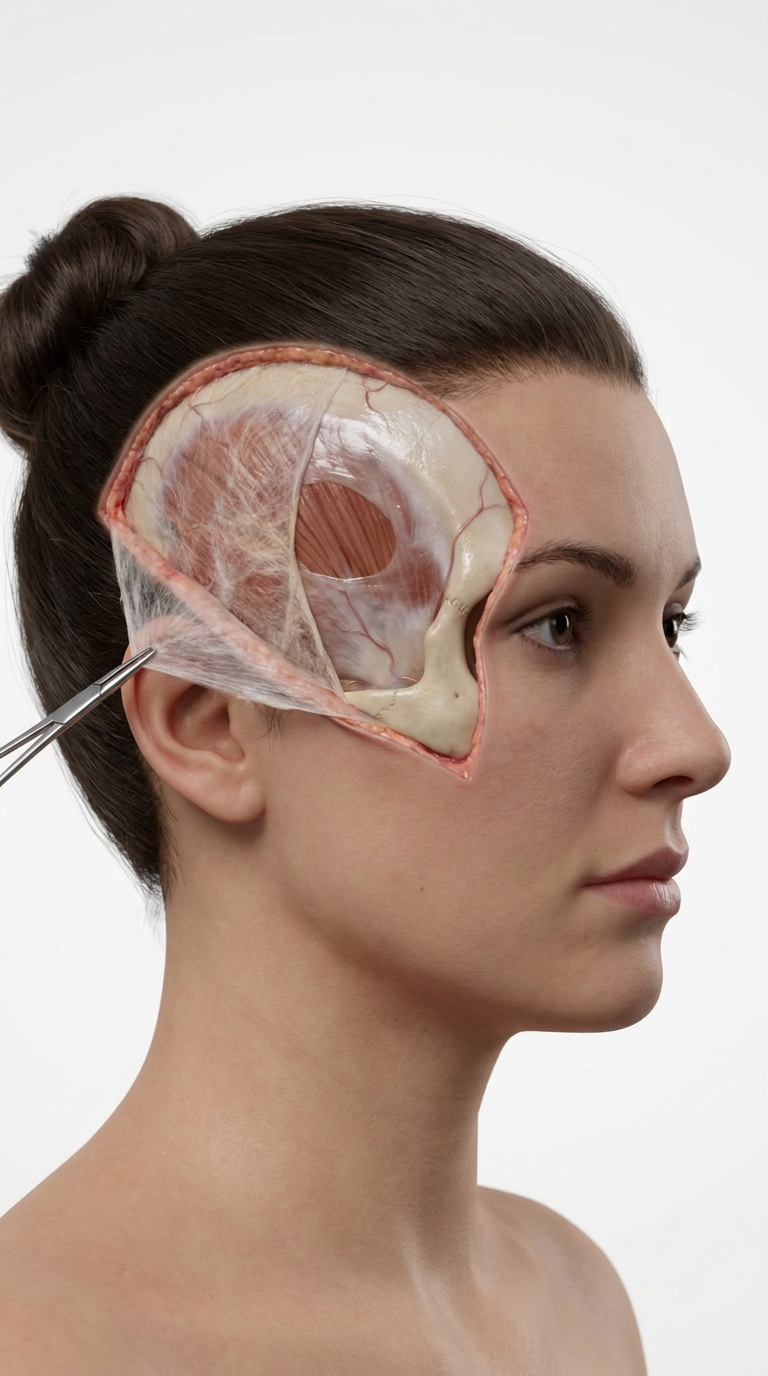
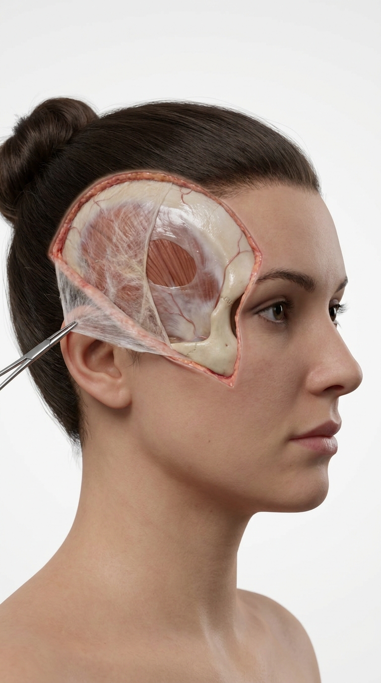
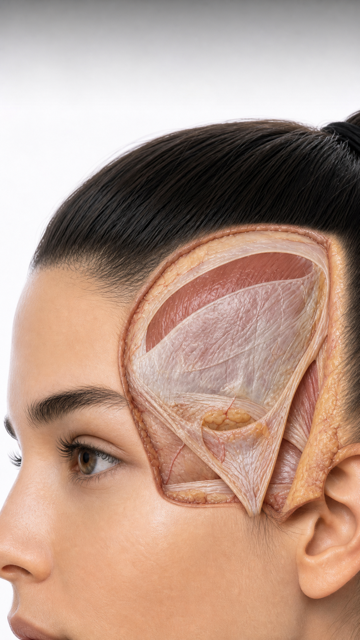
Art style: Cinematic Realistic. Create an original 5 second vertical medical anatomy animation in a 9:16 format. The video must be a newly designed, high-end 3D clinical anatomy visualization rather than footage of a real surgical procedure. Use a completely fictional adult female anatomical model with an original face, hairstyle, head proportions, skin tone, camera angle, dissection geometry, and graphic composition. Show the patient in a stable three-quarter lateral profile against a seamless high-key white studio background. The hair is smoothly retracted superiorly and posteriorly, leaving the temporal and preauricular regions clearly visible. Present one temporal region as a clean, educational anatomical dissection. Create an original curved surgical window extending from the lateral forehead toward the preauricular region, but do not reproduce the shape or borders of any reference image. The skin and subcutaneous tissue are gently reflected at the margins. The superficial temporal fascia, also called the temporoparietal fascia, is elevated as a thin, pliable fascial flap and reflected posteroinferiorly. A small atraumatic surgical forceps holds the distal edge of the flap near the preauricular area. Clearly demonstrate the following anatomical structures: – skin and subcutaneous tissue at the margins of the dissection; – reflected superficial temporal fascia; – loose areolar tissue beneath the superficial fascia; – smooth, pale, slightly glistening deep temporal fascia; – reddish-brown temporalis muscle fibres visible through a separate limited opening in the deep fascia; – superior border of the zygomatic arch at the inferior part of the field; – lateral orbital rim and frontozygomatic region as orientation landmarks; – subtle, anatomically plausible superficial vessels without active bleeding. Maintain the correct anatomical layer sequence: skin and subcutaneous tissue → superficial temporal fascia / temporoparietal fascia → loose areolar tissue → deep temporal fascia → temporalis muscle. Do not merge the superficial temporal fascia with the deep temporal fascia. The loose areolar plane must remain clearly visible as a distinct, delicate intermediate layer. The reflected fascial flap should appear thin, fibrous, slightly translucent, and mechanically plausible. The deep temporal fascia should remain smooth, continuous, and firmly attached over the temporalis muscle. The temporalis muscle fibres should follow a realistic fan-shaped orientation. SHOT SEQUENCE 0–3 seconds: Begin with a close macro view of the temporal dissection. Perform a slow diagonal camera glide from the anterosuperior margin toward the preauricular region. Emphasize the contrast between the thin reflected superficial temporal fascia, the loose areolar plane, and the smooth deep temporal fascia. 3–6 seconds: Slowly dolly backward to reveal the fictional patient’s forehead, eyebrow, lateral orbital rim, eye, cheekbone, ear, and the full extent of the temporal dissection. The head remains completely still. Only the camera moves. 6–9 seconds: Perform a controlled push-in toward the lower temporal and preauricular region. Use subtle parallax to demonstrate the depth relationship between the fascial flap, loose areolar tissue, deep temporal fascia, temporalis muscle, and zygomatic arch. 9–12 seconds: Hold the camera in a slightly oblique close-up. Sequentially fade in thin dark-grey leader lines and small clean sans-serif anatomical labels. Use a completely original label arrangement. Place labels outside the operative field with sufficient spacing and no overlapping lines. Display these labels: Superficial temporal fascia Loose areolar tissue Deep temporal fascia Temporalis muscle Zygomatic arch VISUAL STYLE Photorealistic contemporary 3D medical visualization; premium postgraduate surgical-atlas quality; anatomically rigorous clinical anatomy; clean educational dissection; natural tissue textures with restrained moisture; no excessive shine; soft neutral studio illumination; high-key white background; minimal shadows; subtle depth of field; smooth stabilized cinematic camera movement; realistic proportions and tissue thickness; restrained ivory, beige, pale pink, fascia-white, and muscle-red colour palette; fine fibrous detail in fascial structures; high spatial consistency throughout the animation. The anatomical model, face, hairstyle, surgical window, tissue flap, instrument position, camera trajectory, lighting design, label typography, leader-line placement, and final composition must be newly created from first principles. Use the reference image only to understand the general educational subject of temporal-region layered anatomy. Do not reproduce its exact framing, facial appearance, surgical borders, flap geometry, instrument angle, vessel pattern, labels, graphic lines, colour grading, watermark position, or visual identity. No imitation of any identifiable medical illustrator, animation studio, educational account, publication, social-media page, or copyrighted anatomical artwork. No narration. No subtitles. No logo. No watermark. No username. No social-media interface. No branding. Render in high resolution at 24 or 30 fps. Use smooth motion, stable anatomy, and consistent tissue layers. Avoid flicker, morphing, duplicated vessels, changing facial features, anatomical deformation, shifting dissection margins, or inconsistent instrument placement. ━━━━━━━━━━━━━━━━━━━━━━━━━━━━━━━━━━━━━━ FINAL STYLE ENFORCEMENT (this is the authoritative rendering directive — it overrides any style wording elsewhere in the prompt): Render this image entirely in "Cinematic Realistic" style. Match the lighting grammar, material response, colour science, level of realism / stylisation, line weight, and shading model typical of "Cinematic Realistic". Keep the subject, composition, camera angle, lighting direction, and colour palette from the description above — but EVERY surface, skin, fabric, material, and environmental element must be rendered in the "Cinematic Realistic" idiom, not in any other medium.
Free to start · Generate videos and images with AI in seconds
More from this creator




More L images
См. все →