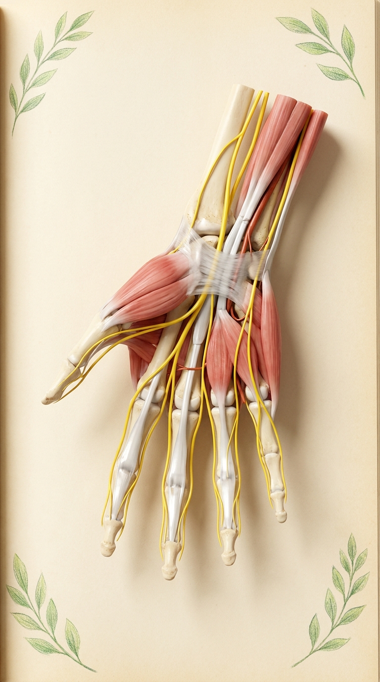
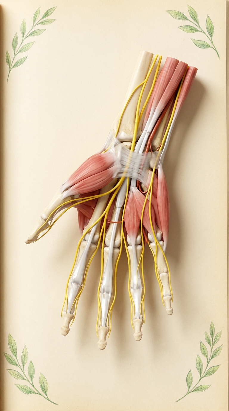
Photorealistic 3D medical anatomy render in the style of a high-end anatomical atlas (like VOKA 3D Anatomy), palmar view of a right human hand and wrist, skin and fat removed. Anatomically accurate: five fingers, natural bone proportions. The median nerve (bright yellow, realistic nerve texture) runs down the forearm, passes through the carpal tunnel beneath the transverse carpal ligament (flexor retinaculum, shown semi-transparent) together with the flexor tendons, then branches into common and proper palmar digital nerves to the thumb, index finger, middle finger and radial half of the ring finger; recurrent motor branch to thenar muscles. The ulnar nerve (yellow) runs on the ulnar side through Guyon's canal next to the ulnar artery and branches to the little finger and ulnar half of the ring finger. Realistic red-pink muscles (thenar and hypothenar eminences, lumbricals), white glossy tendons, ivory bones visible at the wrist. Soft warm studio light, gentle shadows, physiological and clean, not gory. Background: warm cream textured paper page of an old anatomical atlas, a few small delicate green leafy sprigs drawn in colored pencil in the corners, generous empty space around the hand for handwritten labels. No text, no letters, no labels, no watermark. Vertical composition.