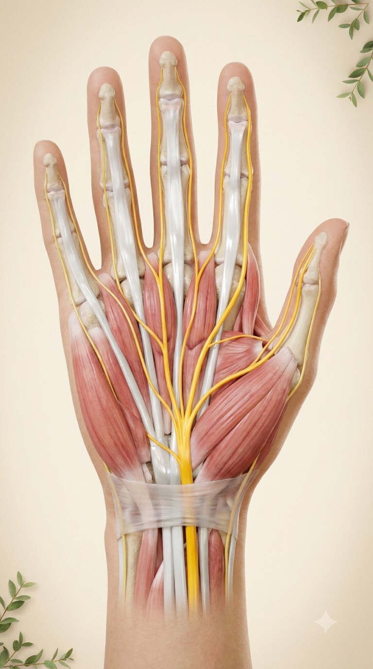
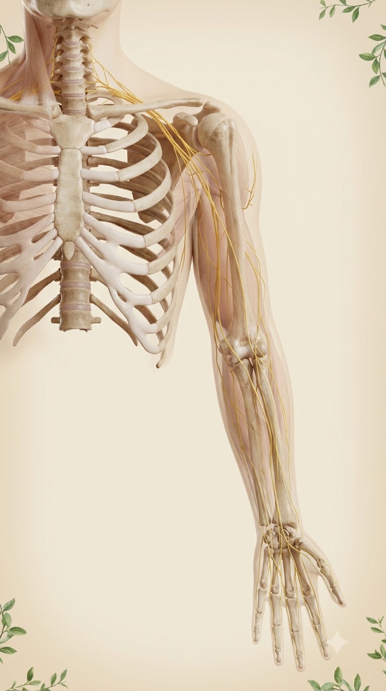
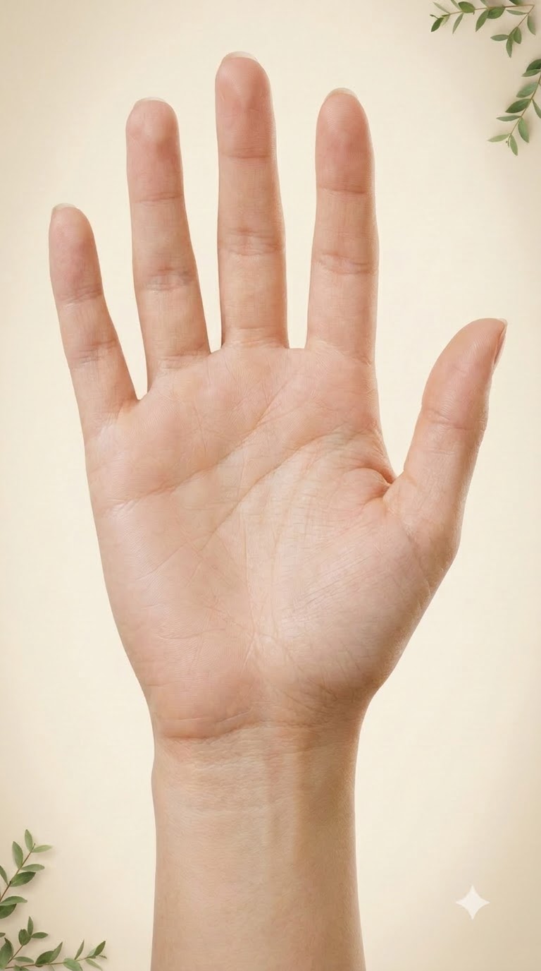
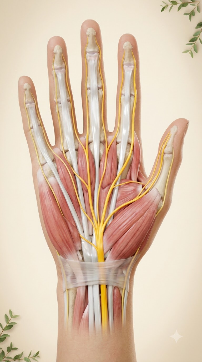
Use the reference image (VOKA 3D anatomy atlas, palmar view of a right hand) as the exact anatomical guide. Create a photorealistic 3D medical anatomy render, quality of a premium 3D anatomy atlas: palmar view of an adult right hand and wrist, palm facing the viewer, fingers pointing up, thumb on the right side of the image. Skin removed. Clearly show the carpal tunnel: the transverse carpal ligament (flexor retinaculum) as a semi-transparent pale band across the wrist; beneath it the flexor tendons and the median nerve (bright warm yellow, realistic nerve fibre texture). Below the ligament the median nerve branches exactly as in anatomy: motor branch to the thenar muscles, and palmar digital nerves running along both sides of the thumb, index finger, middle finger and the thumb-side half of the ring finger. The ulnar nerve (yellow, thinner) runs on the little-finger side to the little finger and the other half of the ring finger. Realistic red-pink muscles (thenar, hypothenar, lumbricals), glossy white tendons, ivory bones visible at the wrist, fingers with their tendons and soft tissue, exactly five fingers. Soft warm studio light, physiological, clean, not gory. Background: plain warm cream paper tone with very soft vignette, a couple of small green leafy sprigs in the corners. No text, no labels, no letters, no watermark. Vertical 9:16, hand centered and filling most of the frame.