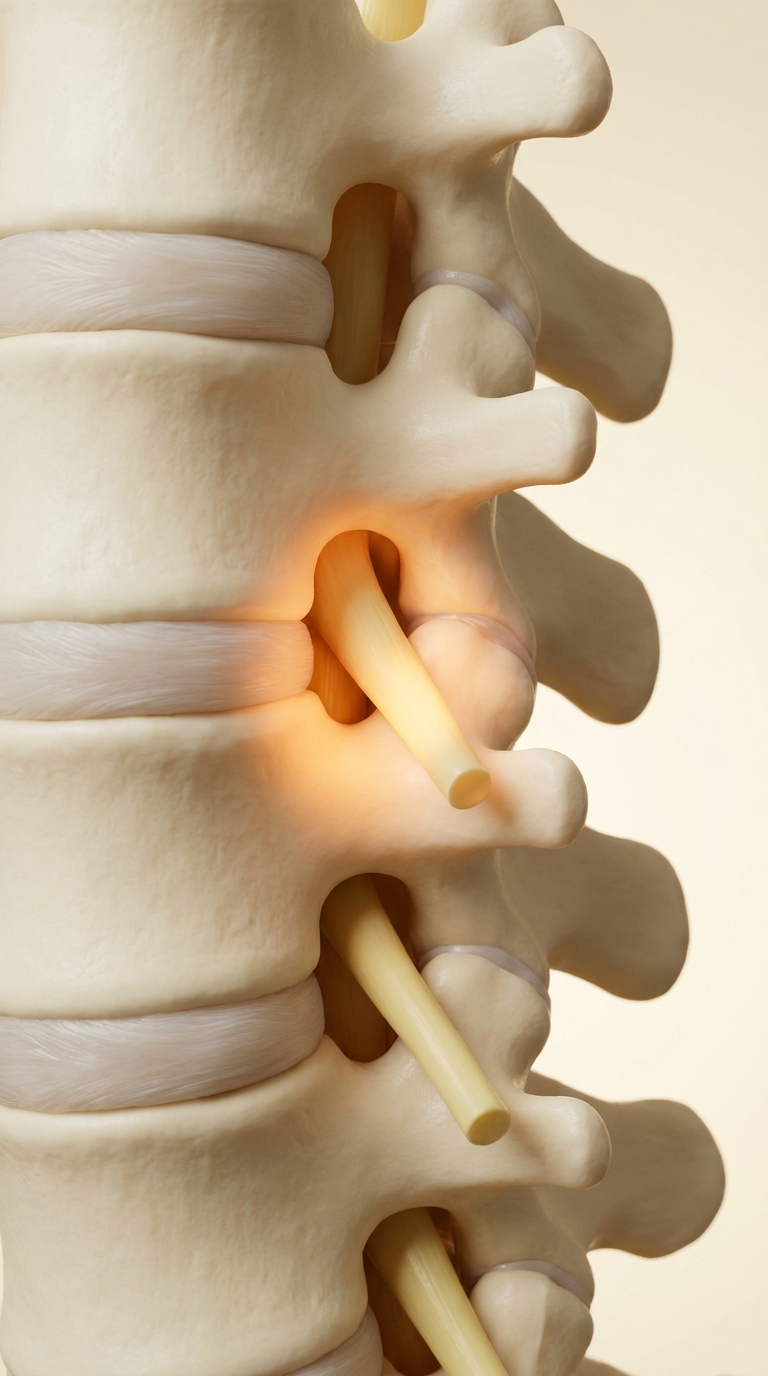
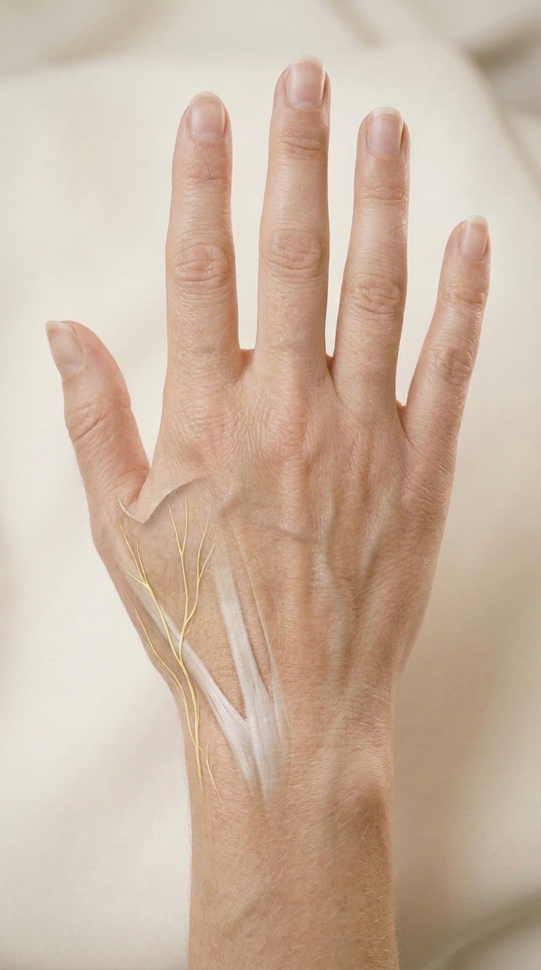
Cervical nerve root, close-up. A realistic medical teaching model of a short segment of the lower cervical spine in true right lateral oblique view on a softly lit warm ivory studio background. Large clear vertebral bodies and pale discs on the anterior side, facet joints and short spinous processes posteriorly, anatomically correct aligned cervical intervertebral foramina between adjacent pedicles. The nearest short pale yellow spinal nerve emerges laterally through its neural foramen, immediately below the pedicle of the vertebra above. One small zone of warm amber illumination at this exit only; bone and nerve remain distinct. Naturally separated adjacent segmental spinal nerves. Do not show a cord through a disc or through a vertebral body. Gentle optical depth, photorealistic 3D educational atlas, restrained contrast, ivory bone and subtle tissue detail, same soft cream palette as a medical photograph of a hand. Portrait 9:16, center the visible nerve exit. No labels, words, arrows, skull, shoulders or clavicles.